3D
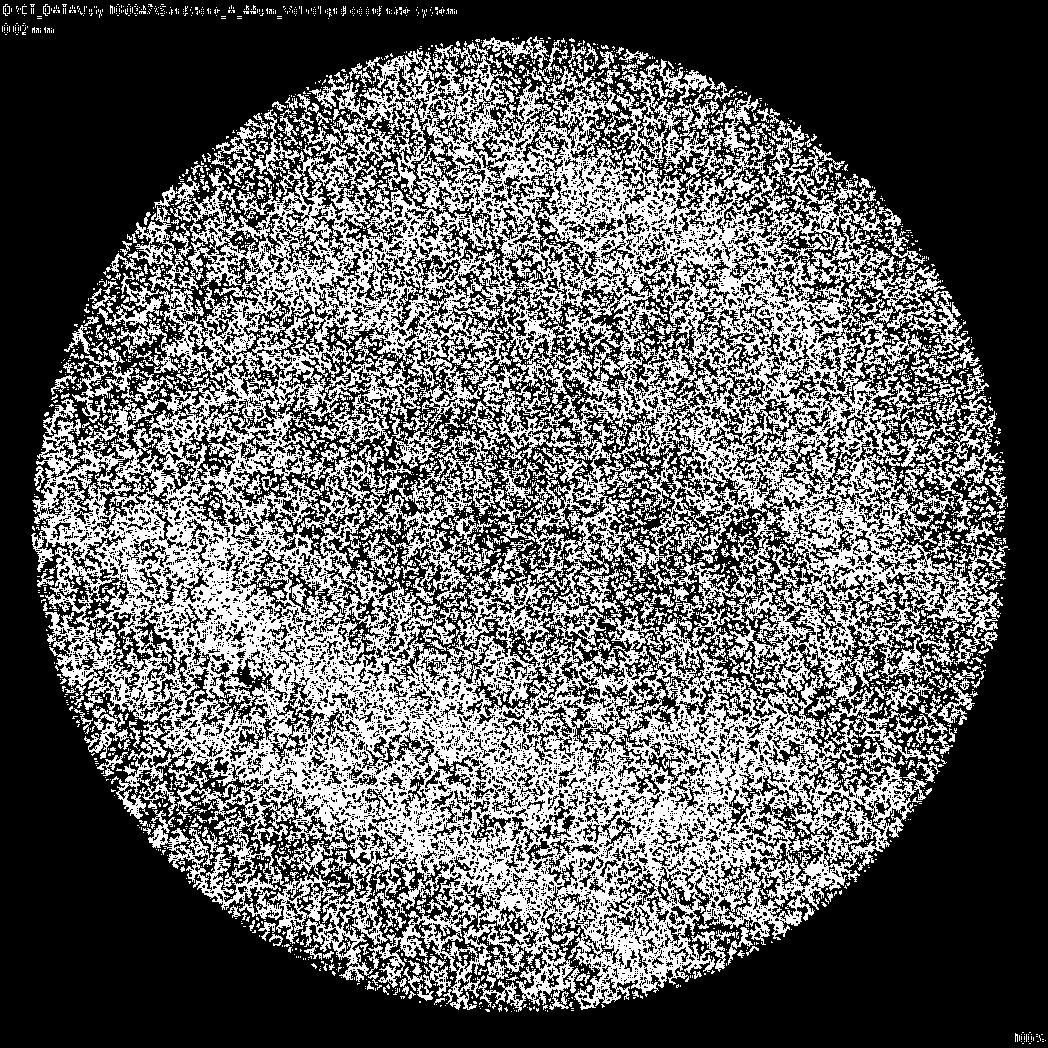
 HI, Im a university student and i'm new to imageJ. For one of my major work, I need to use imageJ to construct a 3D pore stucture of a sandstone sample such as ars.els-cdn.com/content/image/1-s2.0-S0167198712000931-gr1.jpg I have already threshold about 300 slice of the CT-scan images. Is it possible to guide me on how to do it? tried putting them into stacks and using 3d-project to come up with something but it doesn't look similar at all. Thanks! |
|
Hi Julian
First of all, import your microtomography slice images as a series to make an image stack. File>Import>Image Sequence... Then binarize it by picking a threshold. The threshold can be applied to all the slices. Image>Adjust>Threshold (I guess you have strong contrast between sandstone matrix and background, which is e.g. water or air) Then try running Purify, then Connectivity: http://bonej.org/purify http://bonej.org/connectivity Your rock structure might be like trabecular bone, in which there is a single connected network of struts. Or else it might have disconnected particles or pore spaces. If there are several particles / pore spaces then use BoneJ's particle analyser I mentioned before which will tell you the Euler characteristic of each particle. Connectivity is calculated from the Euler characteristic and assumes a single particle with no cavities is in the image. Connectivity can be interpreted as the number of connected branches in a structure. Connectivity density is just that divided by the volume of your sample. Don't bother with 3D Project - that will make a 2D image from your 3D data. Michael ------------------------ <http://imagej.1557.n6.nabble.com/file/n5001765/Sandstone-A_XY_000.jpg> HI, Im a university student and i'm new to imageJ. For one of my major work, I need to use imageJ to construct a 3D pore stucture of a sandstone sample such as ars.els-cdn.com/content/image/1-s2.0-S0167198712000931-gr1.jpg I have already threshold about 300 slice of the CT-scan images. Is it possible to guide me on how to do it? tried putting them into stacks and using 3d-project to come up with something but it doesn't look similar at all. Thanks! [http://www.rvc.ac.uk/cf_images/button_rvc.png]<http://www.rvc.ac.uk> [http://www.rvc.ac.uk/cf_images/button_twitter.png] <http://twitter.com/RoyalVetCollege> [http://www.rvc.ac.uk/cf_images/button_facebook.png] <http://www.facebook.com/theRVC> This message, together with any attachments, is intended for the stated addressee(s) only and may contain privileged or confidential information. Any views or opinions presented are solely those of the author and do not necessarily represent those of the Royal Veterinary College (RVC). If you are not the intended recipient, please notify the sender and be advised that you have received this message in error and that any use, dissemination, forwarding, printing, or copying is strictly prohibited. Unless stated expressly in this email, this email does not create, form part of, or vary any contractual or unilateral obligation. Email communication cannot be guaranteed to be secure or error free as information could be intercepted, corrupted, amended, lost, destroyed, incomplete or contain viruses. Therefore, we do not accept liability for any such matters or their consequences. Communication with us by email will be taken as acceptance of the risks inherent in doing so. -- ImageJ mailing list: http://imagej.nih.gov/ij/list.html |
«
Return to ImageJ
|
1 view|%1 views
| Free forum by Nabble | Edit this page |

