Hello:
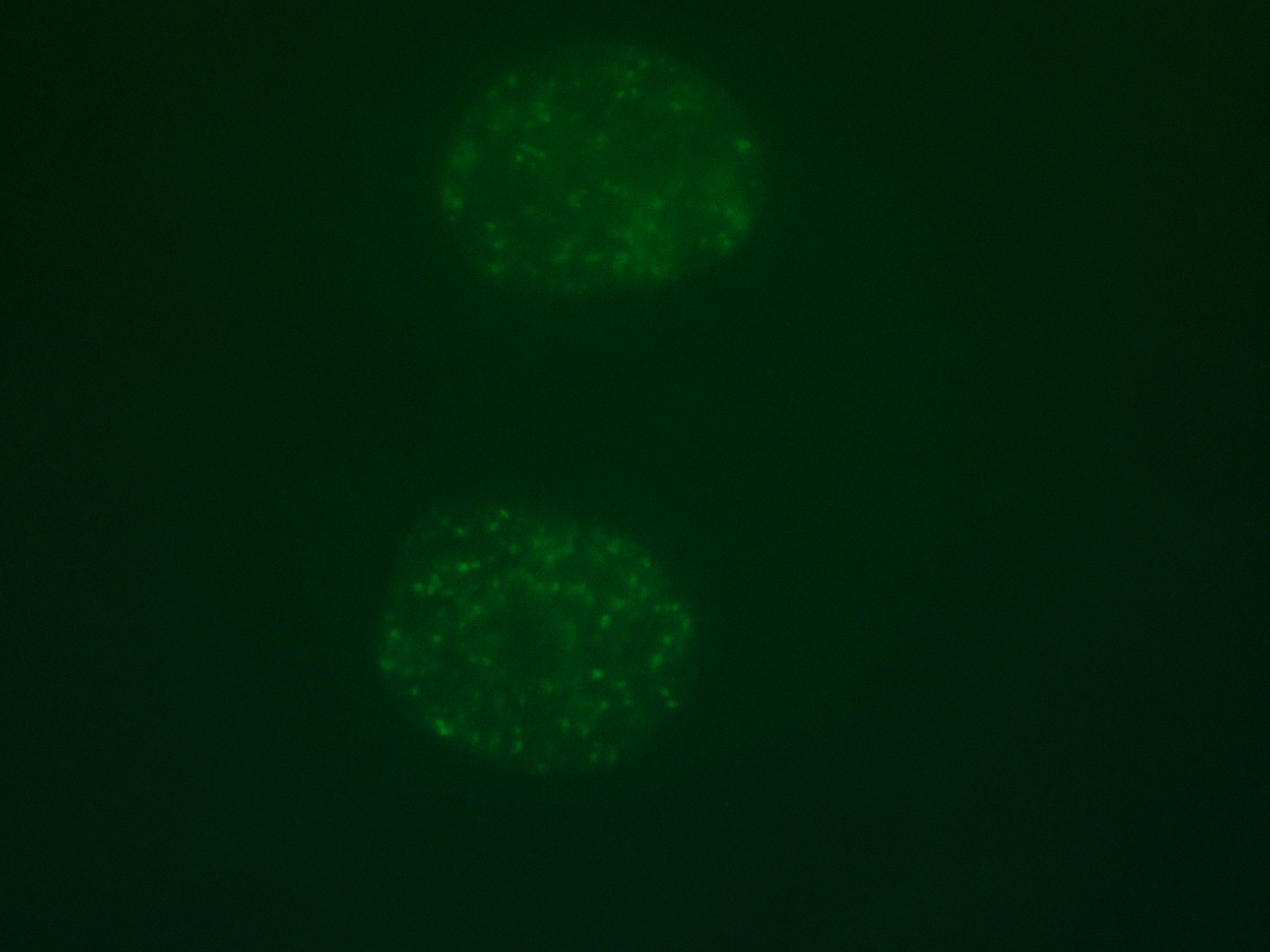
I did immunostaining with r-H2AX fluorescent stain for 293T and MCF10A cell lines. And I took the images with fluorescent microscope in our lab. Please see the attached pictures. I have tried to use following methods to count the foci in each cell
1. adjust bright/contrast
2. subtract background
3. Use color threshold to adjust and cover most foci area (sometimes the image is kinda blur and hard to use this)
4. Binary (convert to mask--fill in holes--watershed)
5. Analysis particles (don't know how to set the size and circus??) --I usually use 100 for size and 2 for circus
I had use this method to count some of my cells and the results didn't turn out as what I expected..
Is there a more efficient way to count the foci through imageJ? How to sharp the image once it is blur...as you can see since my picture is only count one plane of the nucleus, there are more dots hiding in the background and kinda distracting...is there a way to make the dots more brighter?
Thank you for answering my questions..