Quantifying Bone Porosity Using Brightfield or Polarized Images?


|
Hello everyone,
I am a second-year Master's-PhD student studying skeletal biology in the Department of Anthropology. My master's thesis project is to quantify the porosity in cross-sections of different bones. These pores are formed either as Haversian canals (tunnels to carry blood vessels through the bone) or as resorption cavities (larger tunnels of bone loss which are either permanent, or will be reconstructed as new Haversian canals). Each pore is contained with an osteon (circular unit of bone). Typically, a microradiograph is taken to quantify such porosity in a cross-section. In this case thresholding to separate the bone from pore is easy, since the microradiograph takes an image similar to an X-ray of the bone. However, my advisor wanted me to find a way to quantify porosity using our normal brightfield or polarized images from a light transmission microscope, to make a "lower tech" protocol available to our department. My main problem is that these pores are often filled with debris - collapsed blood vessels, fat, embedding material, and so on. This debris is darker than most areas of the bone. So in theory, it should be easy to separate light colored bone from dark colored pore. However, there are some patches of bone that are a similar color of brown to the pores. Therefore, when I am thresholding the image, both the pores and some patches of similarly-colored bone are selected. I have tried all sorts of thresholding methods from ImageJ and Fiji to try to select the pores and not the bone: Manual Thresholding, Automatic Thresholding, Robust Automatic Threshold Selection, TopHat methods, enhancing contrast using various tools, and so on. Here is an example of the original image of a radius cross-section. 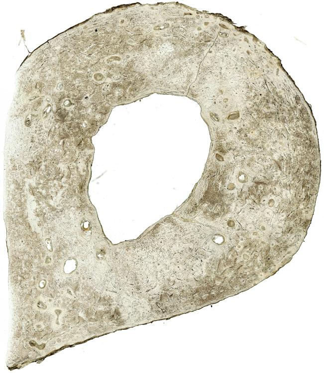 Now I have "cut out" the radius using a digital tablet and then used Enhance Contrast. In this case, some of the pores are clear enough to let the light shine through, so I have already selected their area using Color Threshold and filled them with "Black" to make all of the pores dark. As a note, it appears I had to convert the image from a TIFF to a JPG and resize them to upload to this forum. 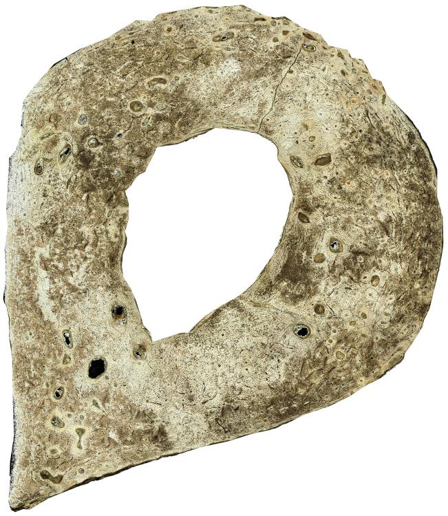 Here is the closest I can get with thresholding. I adjust Brightness/Contrast, convert to 16-bit grayscale, and set the threshold manually using Adjust Threshold. Then I Make Binary so that I can use the command Fill Holes. Playing around with the Binary options and with Graph Cut or Filters sometimes achieves similar results, although nothing significantly better. Finally, I remove the larger patches of bone and smaller holes (for bone cells called osteocytes) that are caught by the threshold using a Morphology plugin called Particles8. This removes patches of bone larger than the larger pore in the picture (which I measure manually) and smaller than an average pore size. Unfortunately, many small jagged patches of bone remain. As you can see, the image contains both real pores (the round spots) and small patches of bone that happen to be a similar color in the original image (the jagged spots): 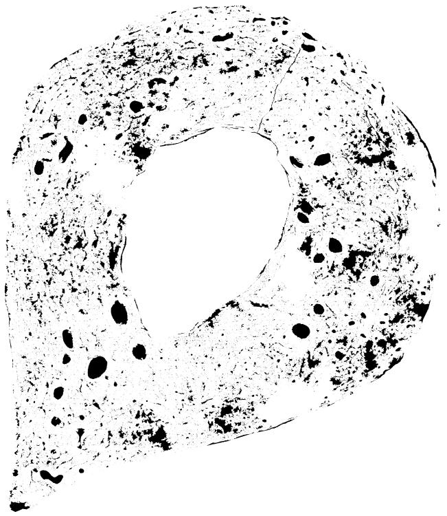 Another option I considered was using a polarized image. In this image, the pores within each osteon (circular unit of bone) are surrounded by white due to the protein content in the bone. Pores are often marked by a characteristic black "X" in the middle of this white circle called a Haversian cross. Here is a picture of the same radius using polarized imaging (slightly off-center from the brightfield image, they were taken on different days): 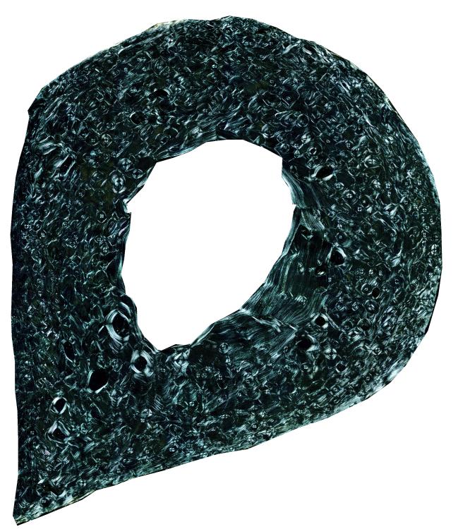 In theory, the white osteons with a Haversian cross would make a target for thresholding. Unfortunately I cannot get this image to be even close to that I obtain from the brightfield using any of the thresholding methods. My main question is: Does anyone know of a way to select the pores and not the bone patches? Even when I set the circularity as high as 0.5, some of the less jagged bone patches are selected and some of the more oblong real pores are NOT selected. I am thinking perhaps of a plugin that detects how "smooth" a pore is around the edge, so as to select the pores only. Another option would be to find a plugin that detects "dark holes," i.e. dark pores surrounded by very bright regions, as my pores appear on the polarized image. Such a plugin appears in Cell Profiler, but that program seems to have trouble running my large TIFF image files. I have not found such a plugin for ImageJ or Fiji. I have also thought of Seeded Region Growing, but for some reason that IJ-Plugin plugin does not work on my system regardless of my updating of Java. I apologize for the length of this message, but I have found the problem very difficult. I have very little experience with image analysis or coding. Thank you for any help you can offer! Sincerely, Mary Cole |
Re: Quantifying Bone Porosity Using Brightfield or Polarized Images?
|
|
Hi Mary,
Maybe very simplistic, but what about using a bone stain first? We often use a von Kossa stain, which stains mineralised tissue black. Other possible stains are anniline blue alizarine red, or picro sirius red for staining the bone. The von Kossa and aniline blue both stain mineral, so need undecalcified bone sections. The other two stains stain the bone matrix. anther easy way to stain the mineralised bone is using calcein blue or green. this will make the mineralised bone brightly blue or green fluorescent. Thresholding the stained bone should be a lot easier. We do this on an almost daily basis for our transgenic mouse bone samples. bye, Rob On 09/07/2013 21:07, Mary Cole wrote: > Hello everyone, > > I am a second-year Master's-PhD student studying skeletal biology in > the Department of Anthropology. My master's thesis project is to quantify > the porosity in cross-sections of different bones. These pores are formed > either as Haversian canals (tunnels to carry blood vessels through the bone) > or as resorption cavities (larger tunnels of bone loss which are either > permanent, or will be reconstructed as new Haversian canals). Each pore is > contained with an osteon (circular unit of bone). > > Typically, a microradiograph is taken to quantify such porosity in a > cross-section. In this case thresholding to separate the bone from pore is > easy, since the microradiograph takes an image similar to an X-ray of the > bone. However, my advisor wanted me to find a way to quantify porosity > using our normal brightfield or polarized images from a light transmission > microscope, to make a "lower tech" protocol available to our department. > > My main problem is that these pores are often filled with debris - > collapsed blood vessels, fat, embedding material, and so on. This debris is > darker than most areas of the bone. So in theory, it should be easy to > separate light colored bone from dark colored pore. However, there are some > patches of bone that are a similar color of brown to the pores. Therefore, > when I am thresholding the image, both the pores and some patches of > similarly-colored bone are selected. > > I have tried all sorts of thresholding methods from ImageJ and Fiji to > try to select the pores and not the bone: Manual Thresholding, Automatic > Thresholding, Robust Automatic Threshold Selection, TopHat methods, > enhancing contrast using various tools, and so on. > > Here is an example of the original image of a radius cross-section. > > <http://imagej.1557.x6.nabble.com/file/n5003863/Original_Image_JPG.jpg> > > Now I have "cut out" the radius using a digital tablet and then used Enhance > Contrast. In this case, some of the pores are clear enough to let the light > shine through, so I have already selected their area using Color Threshold > and filled them with "Black" to make all of the pores dark. As a note, it > appears I had to convert the image from a TIFF to a JPG and resize them to > upload to this forum. > > <http://imagej.1557.x6.nabble.com/file/n5003863/White_Thresholded_and_Histogram_Equalized_JPG.jpg> > > Here is the closest I can get with thresholding. I adjust > Brightness/Contrast, convert to 16-bit grayscale, and set the threshold > manually using Adjust Threshold. Then I Make Binary so that I can use the > command Fill Holes. Playing around with the Binary options and with Graph > Cut or Filters sometimes achieves similar results, although nothing > significantly better. Finally, I remove the larger patches of bone and > smaller holes (for bone cells called osteocytes) that are caught by the > threshold using a Morphology plugin called Particles8. This removes patches > of bone larger than the larger pore in the picture (which I measure > manually) and smaller than an average pore size. Unfortunately, many small > jagged patches of bone remain. > > As you can see, the image contains both real pores (the round spots) and > small patches of bone that happen to be a similar color in the original > image (the jagged spots): > > <http://imagej.1557.x6.nabble.com/file/n5003863/Thresholded_Image_JPG.jpg> > > Another option I considered was using a polarized image. In this image, the > pores within each osteon (circular unit of bone) are surrounded by white due > to the protein content in the bone. Pores are often marked by a > characteristic black "X" in the middle of this white circle called a > Haversian cross. > > Here is a picture of the same radius using polarized imaging (slightly > off-center from the brightfield image, they were taken on different days): > > <http://imagej.1557.x6.nabble.com/file/n5003863/Polarized_Image_JPG.jpg> > > In theory, the white osteons with a Haversian cross would make a target for > thresholding. Unfortunately I cannot get this image to be even close to that > I obtain from the brightfield using any of the thresholding methods. > > My main question is: Does anyone know of a way to select the pores and not > the bone patches? Even when I set the circularity as high as 0.5, some of > the less jagged bone patches are selected and some of the more oblong real > pores are NOT selected. I am thinking perhaps of a plugin that detects how > "smooth" a pore is around the edge, so as to select the pores only. > > Another option would be to find a plugin that detects "dark holes," i.e. > dark pores surrounded by very bright regions, as my pores appear on the > polarized image. Such a plugin appears in Cell Profiler, but that program > seems to have trouble running my large TIFF image files. I have not found > such a plugin for ImageJ or Fiji. > > I have also thought of Seeded Region Growing, but for some reason that > IJ-Plugin plugin does not work on my system regardless of my updating of > Java. > > I apologize for the length of this message, but I have found the problem > very difficult. I have very little experience with image analysis or coding. > Thank you for any help you can offer! > > Sincerely, > Mary Cole > > > > > > > -- > View this message in context: http://imagej.1557.x6.nabble.com/Quantifying-Bone-Porosity-Using-Brightfield-or-Polarized-Images-tp5003863.html > Sent from the ImageJ mailing list archive at Nabble.com. > > -- > ImageJ mailing list: http://imagej.nih.gov/ij/list.html > -- _____________________________ Dr. Rob van 't Hof Reader Centre for Molecular Medicine MRC IGMM University of Edinburgh Western General Hospital Crewe Road, Edinburgh EH4 2XU United Kingdom Phone: (+44)-131-6511031 email: [hidden email] _____________________________ -- ImageJ mailing list: http://imagej.nih.gov/ij/list.html |
|
|
Hello,
Thank you for your reply! I really wish that I could stain the bone samples. Unfortunately, I am working with a collection of cross-sections of bone that were mounted onto slides without staining back in the 1970s. I also thought to use a red filter in the microscope to darken the pores (as someone suggested for a similar problem on this listserv), but our lab apparently does not have any colored filters either. Our lab has no funding for master's thesis projects as they are supposed to be somewhat small scale, so I unfortunately cannot obtain more samples to stain at this time. I do have access to ten images of rib cross-sections stained with Basic Fuchsin, so I will try my method on those images and see if it works. Sincerely, Mary Cole |
Re: Quantifying Bone Porosity Using Brightfield or Polarized Images?
|
|
If using a colour camera can use the ratio's of colours as an equivalent of a filter, (you can even try gaffer taping a camera phone to the eye piece). Borrow a fluorescent microscope as bone is naturally quite fluorescent under blue light (488nm).
Unfortunately for you the system sucks, are the sections even the same thickness? I'm sure it might be possible to calibrate one to the other but really without doing a proper analysis in various thicknesses it seems a typical half arsed masters fob off project to me. (not helpful, sorry) Kenton On 10 Jul 2013, at 02:21, Mary Cole wrote: > Hello, > > Thank you for your reply! I really wish that I could stain the bone > samples. Unfortunately, I am working with a collection of cross-sections of > bone that were mounted onto slides without staining back in the 1970s. I > also thought to use a red filter in the microscope to darken the pores (as > someone suggested for a similar problem on this listserv), but our lab > apparently does not have any colored filters either. > > Our lab has no funding for master's thesis projects as they are supposed to > be somewhat small scale, so I unfortunately cannot obtain more samples to > stain at this time. I do have access to ten images of rib cross-sections > stained with Basic Fuchsin, so I will try my method on those images and see > if it works. > > Sincerely, > Mary Cole > > > > -- > View this message in context: http://imagej.1557.x6.nabble.com/Quantifying-Bone-Porosity-Using-Brightfield-or-Polarized-Images-tp5003863p5003867.html > Sent from the ImageJ mailing list archive at Nabble.com. > > -- > ImageJ mailing list: http://imagej.nih.gov/ij/list.html -- ImageJ mailing list: http://imagej.nih.gov/ij/list.html |
«
Return to ImageJ
|
1 view|%1 views
| Free forum by Nabble | Edit this page |