Hi,
We are trying measure amount of fluorescent glucose uptake in cells with or without the presence of insulin.
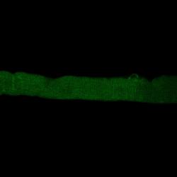
What we see is, without insulin, the fiber has a diffuse green fluorescent haze.
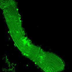
With insulin, bright green spots (various sizes) appear.
We have tried using a Integrated density measurement (with corrected total cell fluorescence), but it does not really reflect what we see.
The measurement of the bright spots in the insulin treated cells seem to be dwarfed by the overall background in the no insulin picture.
So we are not getting much of a difference in overall fluorescence when we use this method.
I have attached two sample images. These are confocal. Care has been taken that every picture has been taken with the same settings. (Yes, these images were pretty good with integrated density, but it is not always that clear. In some experiments background fluorescence is much higher or some fibers have a fewer number of spots)
I am not very literate in the use of ImageJ, so simple directions and advice will be appreciated.
Thank You,
Nancy

