Removing Ring Artefacts
|
Hello All,
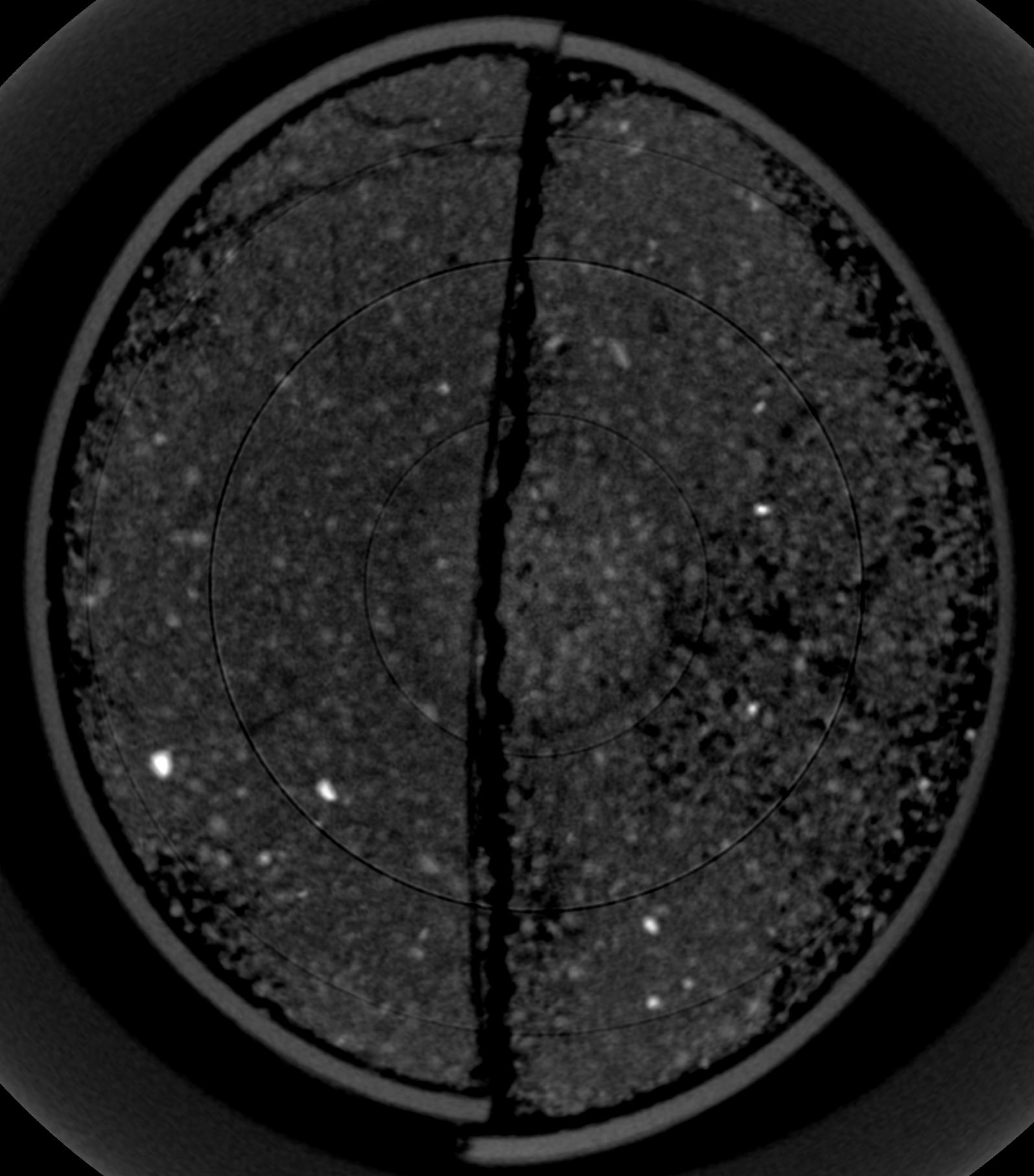
I am currently working on X-Ray CT images of soil within plastic columns (which have been spilt in half with mesh). See the attached image for an example. I am having problems removing the ring artefacts present within my images that will effect the analysis of pore spaces present within these soil columns. I have checked the archives of this mailing group and the plugins page for ImageJ and cannot find any evident solution to the ring artefact problem. I know that this is a common problem, so I hope that there is a solution out there someone which I have simply missed on my research. Any help you can offer will be appreciated very much. Thanks, Sarah 
|
|
>Hello All,
> >I am currently working on X-Ray CT images of soil within plastic columns >(which have been spilt in half with mesh). See the attached image for an >example. I am having problems removing the ring artefacts present within my >images that will effect the analysis of pore spaces present within these >soil columns. I have checked the archives of this mailing group and the >plugins page for ImageJ and cannot find any evident solution to the ring >artefact problem. I know that this is a common problem, so I hope that there >is a solution out there someone which I have simply missed on my research. > >Any help you can offer will be appreciated very much. > >Thanks, >Sarah I'm quite sure that those rings are not artefacts of CT image formation. So could you please be more specific and point us to the source of your opinion: "I know that this is a common problem..." Best -- Herbie ------------------------ <http://www.gluender.de> |
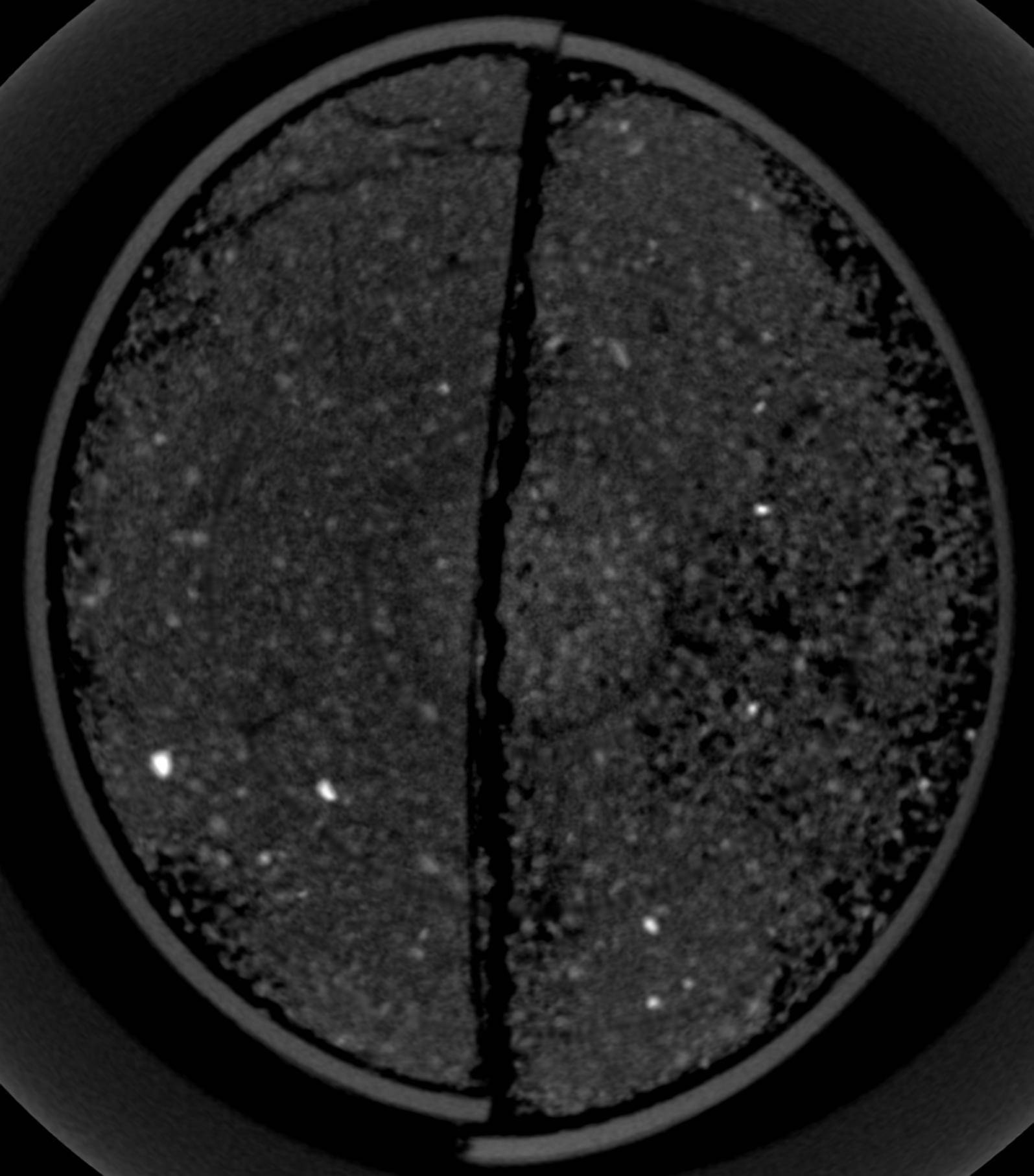
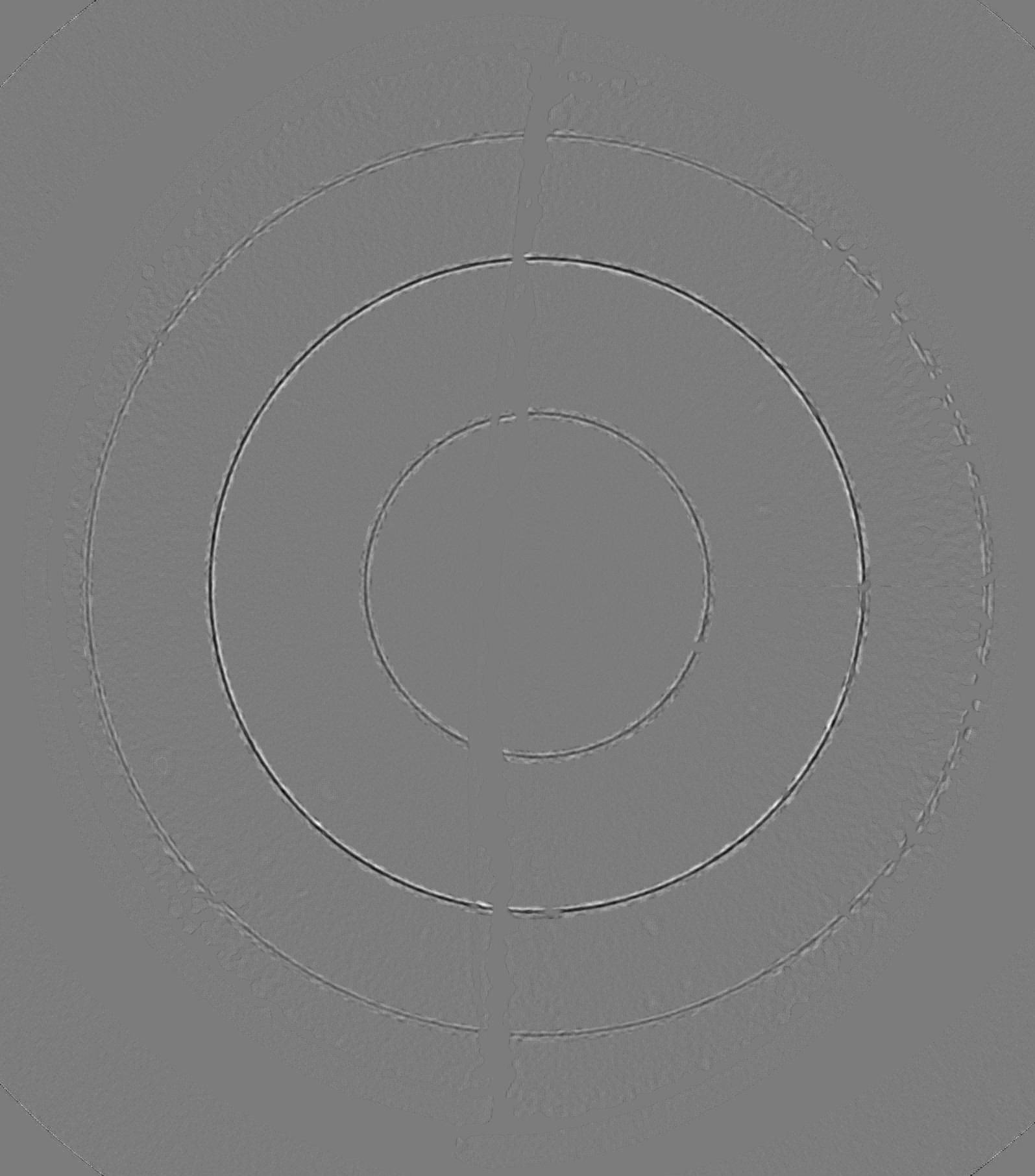
Ring artifacts are common on all real world CT image systems. They correspond for example to defects of the detector. The artifact will be at a fixed position on the projections and thus form a circle (360 degrees scan) or half circle (180 degrees scan) on the tomographic slice. The best solution is to avoid these artifacts at the source or to correct on the projections. In case only reconstructed slices are available several methods are proposed in the literature. A good starting point in ImageJ could be the polar transform and its inverse: http://rsbweb.nih.gov/ij/plugins/polar-transformer.html After transforming to polar coordinates, the rings will appear as vertical lines. The basic idea is that it will be easier to separate these vertical lines from the object information. After filtering the vertical lines in some way, it is sufficient to go back to cartesian coordinates. I made a quick trial on the soil image. As filtering I used an horizontal average on a (manual !) selection of each vertical line. The main steps are recorded below. Maybe this can help as a starting point. Peter Some results: Corrected image:  Difference:  Polar transform (360 angles instead of the 1500 used):  run("Polar Transformer", "method=Polar degrees=360 number=1500 center=827.4 center=902.4"); FILTER ON MANUAL SELECTION AROUND VERTICAL LINE run("Convolve...", "text1=[1 1 1 1 1 1 1 1 1\\n] normalize"); run("Polar Transformer", "method=Cartesian degrees=360 for center=827.4 center=902.4"); run("Specify...", "width=1595 height=1813 x=0 y=0"); run("Crop"); |
|
In reply to this post by Sarah Martin
Hi,
If your rings are concentric around the center of the CT system (isocenter) then they are due to poor calibrations. In 3rd generation CT scanners the detectors and source rotate around the isocenter. If a detector is poorly calibrated the rays from the source to that detector will be incorrect. Those rays always pass at a constant distance from the isocenter. There will be a "ring" at that distance. Jon Sarah Martin wrote: > Hello All, > > I am currently working on X-Ray CT images of soil within plastic columns > (which have been spilt in half with mesh). See the attached image for an > example. I am having problems removing the ring artefacts present within my > images that will effect the analysis of pore spaces present within these > soil columns. I have checked the archives of this mailing group and the > plugins page for ImageJ and cannot find any evident solution to the ring > artefact problem. I know that this is a common problem, so I hope that there > is a solution out there someone which I have simply missed on my research. > > Any help you can offer will be appreciated very much. > > Thanks, > Sarah > > http://n2.nabble.com/file/n1131486/Slice0005.jpg |
|
In reply to this post by pcloetens
>Gluender-2 wrote:
>> >>>Hello All, >>> >>>I am currently working on X-Ray CT images of soil within plastic columns >>>(which have been spilt in half with mesh). See the attached image for an >>>example. I am having problems removing the ring artefacts present within >my >>>images that will effect the analysis of pore spaces present within these >>>soil columns. I have checked the archives of this mailing group and the >>>plugins page for ImageJ and cannot find any evident solution to the ring >>>artefact problem. I know that this is a common problem, so I hope that >there >>>is a solution out there someone which I have simply missed on my research. >>> >>>Any help you can offer will be appreciated very much. >>> >>>Thanks, >>>Sarah >> >> >> I'm quite sure that those rings are not artefacts of CT image >> formation. So could you please be more specific and point us to the >> source of your opinion: >> >> "I know that this is a common problem..." >> > > Best > > Herbie > >Ring artifacts are common on all real world CT image systems. They >correspond for example to defects of the detector. The artifact will be at a >fixed position on the projections and thus form a circle (360 degrees scan) >or half circle (180 degrees scan) on the tomographic slice. The best >solution is to avoid these artifacts at the source or to correct on the >projections. [...] Thanks, this is correct but I use to assume that scientists work with fully functional and adjusted equipment. Again an example where image processing is to be applied to remedy problems with image acquisition... Best -- Herbie ------------------------ <http://www.gluender.de> |
Hi, Thanks for the pointers/help so far! These images were obtained for me by someone else, so I was unaware of any problems until I opened the images, otherwise a correction could have been made earlier on. I did find out that the CT scanner (used to collect these images) had its detectors changed after my samples had been scanned, so obivously there were some problems/ or inadequate adjustments which have raised this issue for me. Regards, Sarah |
«
Return to ImageJ
|
1 view|%1 views
| Free forum by Nabble | Edit this page |

