Hi Ziyi,
it depends on the image type whether 'Fit Polynomial' or 'Subtract
Background' are better. Unfortunately, I can't see your image
('maximum downloads exceeded' error).
Subtract Background requires that the object are EITHER brighter OR
darker than the background. For dark objects, there should be
nothing in the image brighter than what you consider background, and
for bright objects, nothing in the image should be darker than the
background.
Fit Polynomial (also the Highpass filter, which subtracts the
Gaussian-blurred image) is based on the average brightness. So, if
you have large or many objects that are on average significantly
brighter or darker than the background, they won't work well.
Fit Polynomial takes the background from the selection only. So, if
you can get an approximate selection or mask with your objects as a
first step (Thresholding, Versatile Wand with gradient detection, or
whatever), this will help you: Create the inverse selection
(Edit>Selection>Make Inverse) and run Fit Polynomial with that
selection (the background).
[P.S: Have I guessed your name correctly from the email address?
Maybe I am an old-fashioned European who prefers to talk to people
with names...]
Michael
________________________________________________________________
On 8 Oct 2011, at 03:56, GloryField wrote:
> Hi Tom and Michael,
>
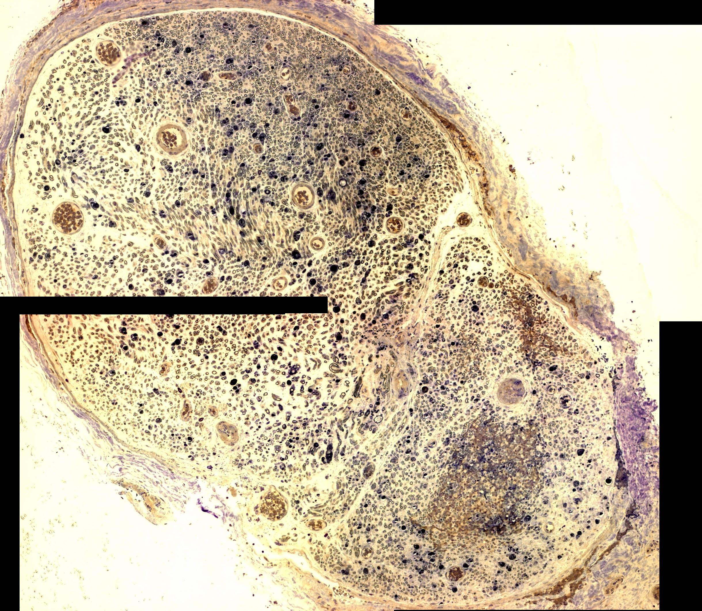
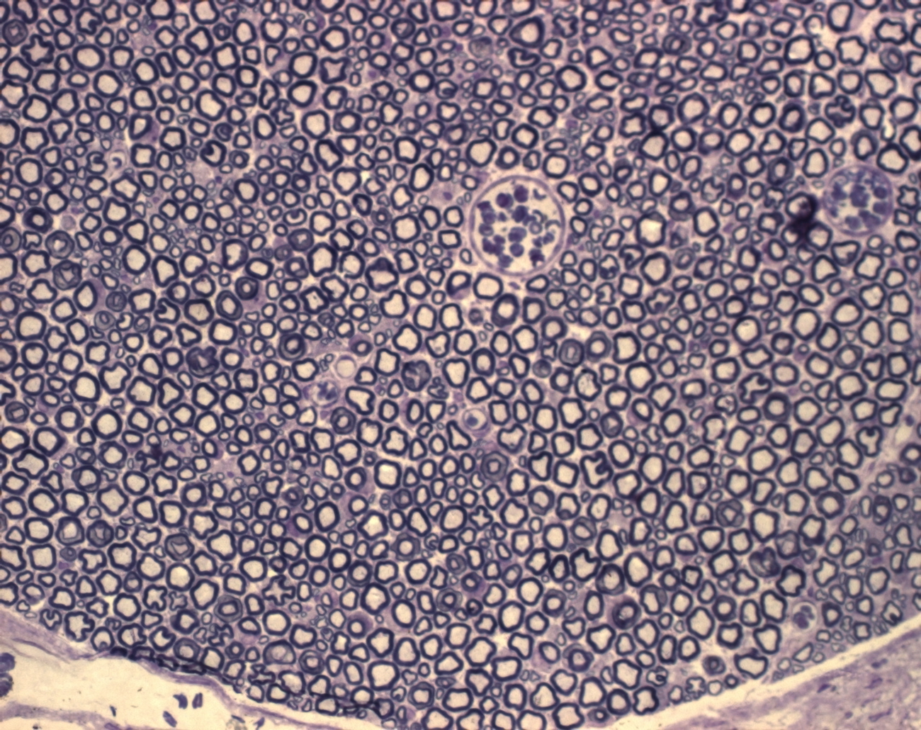
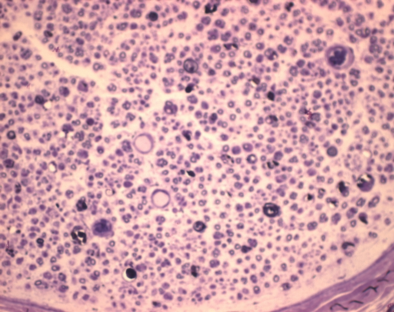
> I'm trying to do something similar, except with axons in a nerve cross
> section, with lots of background.
> I have been using the default "Subtract Background" function within
> ImageJ,
> would the Fit Polynomial work better? I'm very new to working with
> ImageJ.
>
> here's a sample image:
>
http://www.swapdrive.com/file.asp?ID=tG+4c
> +dQUV2uaBUzGE9QXIqvYD6NpPr0zU3HMp4maEzHuo55/04pqQ==url
>
> I'm trying to measure the average area of each of the axons within the
> nerve, including the myelin sheath. What I did so far is:
>
> Process>Subtract Background>5 pixels
> Process>Binary>Make Binary
> Analyze>Analyze Particles>Size 4-350, circularity 0.25-1.0, show
> outlines,
> include holes
>
> I chose 350 as max size because that allows me to eliminate blood
> vessels
> within the nerve.
> I arbitrarily chose 0.25 circularity because I didn't know what
> else to
> pick.
>
> Doing this gave me an average size of 50.668 pixels. I will put a
> slide
> with a length marker under microscope with same magnification so I
> can use
> "Analyze > Set Scale" and get values in micrometers.
>
> Do you guys think this would be an OK way of measuring the axon cross
> section area?
>
> Thank you again!
>
> --
> View this message in context:
http://imagej.588099.n2.nabble.com/
> Separating-cells-close-to-each-other-tp6869352p6871669.html
> Sent from the ImageJ mailing list archive at Nabble.com.