Using ImageJ for dental radiograph measurements
|
Hello all
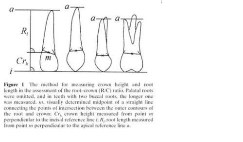
Sorry if this query is extremely basic, but I have spent a long time searching the archives and reading the instruction manuals and can't find a clear answer, so I thought someone here may be able to quickly solve my problem! I need to measure tooth dimensions from digitised panoramic radiographs. The method I'm using looks like this:  Is there a simple way in ImageJ to draw the perpendicular lines on the image and then record the measurements to calculate the ratio of crown length to root length? I don't want the perpendicular lines to remain on the image after I've done the measurement as I need to do each tooth, and they are quite close together on a panoramic radiograph. Hoping that someone is able to help and apologies if my query is extremely basic! Thanks in advance ~ Minnie |
|
I am still new to ImageJ, so hopefully others can offer alternative suggestions.
*1) Set Global Scale 1a) Open an image with a scale bar, and draw a delection line over it (click shift to create a perfectly level line). 1b) Go to Analyze --> Set Scale. In the Set Scale window the length of the line, in pixels, will be displayed. Type the known distance and units of measure. Measurements will now be shown using these settings. If the pixel:length relationship is known from a previous measurement you may directly type this information in the Set Scale window. 1c) Alternatively, if one already knows the pixels/unit of length, such as px/um, then one may directly enter that value in the "Distance in Pixels." One also needs to enter 1 as the "Known Distance", and the desired "Unit of Length" such as micron. 1d) Check 'global' to apply this scale to other image frames. 1e) When opening new images make sure to not have the global scale disabled! Source: http://rsbweb.nih.gov/ij/docs/menus/analyze.html#scale *2) Using the ROI (Region of Interest) Manager 2a) Open the ROI Manager: Analyze --> Tools --> ROI Manager 2b) Make a selection (explained below) and click "add" on the ROI Manager or use the keyboard shortcut "t" to save the selection to the ROI Manager. 2c) To update a selection (if you made a mistake), simply select the correct ROI in your image and then highlight the correct ROI you want to update in the ROI Manger and select "update" 2d) To delete an ROI select it in the ROI Manager and select "delete". 2e) Select "show all" to view all the selections, select "show all" again to hide the selections. 2f) Once all your selections are complete, click "deselect" in the ROI Manager (to make sure no single ROI is selected in the manager such that they are "all" selected) and select "save" and name the file with "- Compressed - Selections" ending such as: 20090922 #1 60x - Compressed - Selections.zip 2g) To show one's selections, simply open the corresponding image and the ROI Manager and select "open" and navigate to the corresponding .zip file containing the selection information. Source: http://rsbweb.nih.gov/ij/docs/menus/analyze.html#manager *3) Creating Selections 3a) Zoom into the selection area using the zoom tool (left click = zoom, right click = zoom out). The zoom level is displayed in the image's title bar. Double click the zoom icon to reset the image's zoom level. There are multiple ways to select a region of interest, such as: 3b) Freehand Selection: Simply freehand a selection 3c) Segmented Line: Click to create a segmented line in which the segments can be "pulled" or "pushed" to modify the selection. 3d) Freehand Line: Similar to the freehand selection tool, but works well on more pixilated boarders. 3e) It is often helpful to view the composite image at the same time such that you can move through the slices to clearly see cells and faint areas on the compressed image. Source: http://rsbweb.nih.gov/ij/docs/tools.html *Note: Hold shift down while making your selection to form a perfectly straight line. *4) Creating Advanced Selections 4a) Advanced selections do not work well for these images since the fluorescence covers many structures of non-interest and is not uniform. See the following resources: * Page 43 of "Image processing and analysis with ImageJ and MRI Cell Image Analyzer.pdf" from Montpellier RIO Imaging by Volker Baecker on 13.05.2009 * Area Measurements and Particle Counting.pdf by Larry Reinking on August 30, 2001 - http://rsbweb.nih.gov/ij/docs/pdfs/examples.pdf * Page 6 of "3D Processing and Analysis with ImageJ.pdf" by Thomas Boudier *5) Set Measurements Properties 5a) To select what measurements will be analyze navigate to: Analyze --> Set Measurements 5b) Uncheck all but "Feret's Diameter" and change "Decimal Places" to 2. Source: http://rsbweb.nih.gov/ij/docs/menus/analyze.html#set Note: Read the source link above and play with other measurement possibilities such as bounding rectangle. *6) Make Measurements 6a) Make sure the image and the selections are open (see step 4g above). 6b) In the ROI Manager select "Measure". Alternatively one could use Analyze --> Measure 6c) To save the results to excel select File --> Save As or Edit --> Select All and Copy. 6d) Perform your ratio measurements in excel. Source: http://rsbweb.nih.gov/ij/docs/menus/analyze.html#manager http://rsbweb.nih.gov/ij/docs/menus/analyze.html#measure -Kalen |
|
In reply to this post by MinnieB
Hello Minnie,
I am working on a plugin to do some of this measurements. It will be posted after some testing. But I have a question: What is the source of the figure in your post? I am working on collecting some theory around those measurements. Kind regards Gerald R. Torgersen Physicist Faculty of Dentistry University of Oslo |
|
Hi there
I'm not sure exactly which figure you're asking about, but the method is taken from this paper: Holtta P, Nystrom M, Evalahti M, Alaluusua S. Root-crown ratios of permanent teeth in a healthy Finnish population assessed from panoramic radiographs. Eur J Orthod. 2004 Oct;26(5):491-7. ~ Minnie Date: Thu, 10 Jun 2010 23:16:07 -0700 From: [hidden email] To: [hidden email] Subject: Re: Using ImageJ for dental radiograph measurements Hello Minnie, I am working on a plugin to do some of this measurements. It will be posted after some testing. But I have a question: What is the source of the figure in your post? I am working on collecting some theory around those measurements. Kind regards Gerald R. Torgersen Physicist Faculty of Dentistry University of Oslo
View message @ http://imagej.588099.n2.nabble.com/Using-ImageJ-for-dental-radiograph-measurements-tp4035332p5166615.html
To unsubscribe from Using ImageJ for dental radiograph measurements, click here. Looking for a hot date? View photos of singles in your area! |
|
In reply to this post by MinnieB
Hey all this information was very helpful for me. I just wanted to know about a good dentist Redondo Beach for my four year old son’s regular checkups. If you know about any good dentist that is easy with kids then please let me know.
|
Re: Using ImageJ for dental radiograph measurements
|
In reply to this post by Kalen K
Thanks alot dear
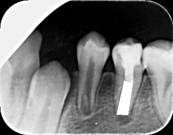
I want to ask a question if you can help me about the use of imageJ for measurements of area from a digital radiograph on meters the area filled with white color  best regards Nazim |
«
Return to ImageJ
|
1 view|%1 views
| Free forum by Nabble | Edit this page |

