Re: Help with surface descriptors!
Posted by Giuseppe Lucarelli on
URL: http://imagej.273.s1.nabble.com/Help-with-surface-descriptors-tp5009390p5009408.html
Hi Charles,
Thank you so much for your reply.
Yes you are right my images were shocking! Here are full size fields of view from my microscope.
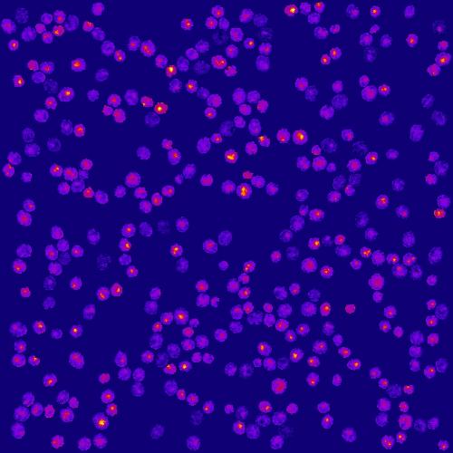
Cells with a relatively central peak:
Cells with irregular/edge peaks or flat:
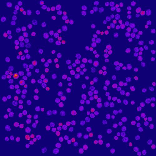
The LUT is fire and the yellow/red areas represent vacuoles when they are relatively central and stand out from the rest of the cell. I have ROIs for every cells in each image and I need to make another ROI for that peaked region given that it has the following criteria:
1. needs to have a minimum intensity of 3 (this is arbitrary for now but I am planning to fix this);
2. The maximum of this peaked region cannot be on the edge of the cell;
3. The peaked region must have a clear "valley" that separates it from the rest of the cell.
4. I need to make a selection of this peaked area and add it as an ROI for downstream measurements (I do this at the moment by finding the peak in the cell and then using the wand tool to grow an area)
I hope this is clear and sorry for wasting your time.
Thanks a lot.
Giuseppe
URL: http://imagej.273.s1.nabble.com/Help-with-surface-descriptors-tp5009390p5009408.html
Hi Charles,
Thank you so much for your reply.
Yes you are right my images were shocking! Here are full size fields of view from my microscope.
Cells with a relatively central peak:
Cells with irregular/edge peaks or flat:
The LUT is fire and the yellow/red areas represent vacuoles when they are relatively central and stand out from the rest of the cell. I have ROIs for every cells in each image and I need to make another ROI for that peaked region given that it has the following criteria:
1. needs to have a minimum intensity of 3 (this is arbitrary for now but I am planning to fix this);
2. The maximum of this peaked region cannot be on the edge of the cell;
3. The peaked region must have a clear "valley" that separates it from the rest of the cell.
4. I need to make a selection of this peaked area and add it as an ROI for downstream measurements (I do this at the moment by finding the peak in the cell and then using the wand tool to grow an area)
I hope this is clear and sorry for wasting your time.
Thanks a lot.
Giuseppe
| Free forum by Nabble | Edit this page |