Hello,
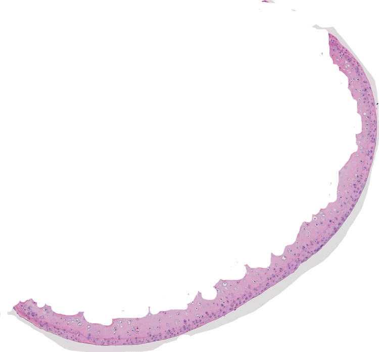
I have H&E stained sagittal cross-sections of mouse knees and I need to count the cells in the femoral cartilage. I cropped the image so that i am only viewing the cartilage and have the Color Deconvolution tool. What is the best way to get nuclei (cell) counts? It seems as though the ITCN plugin is very helpful and I can standardize the threshold and other settings.
I found that it's difficult to get a very clear image so that ITCN can count the nuclei. Would you suggest converting to 8-bit and adjusting contrast? I have attached an image of the femoral cartilage with H&E.
Any suggestions would be of much help! If you have a picture of an optimal contrast setting for cell counting, could you please attach it?
Thank you,
Merissa