Detecting dark spots (dead cells)

Previous Topic Next Topic
 
classic Classic list List threaded Threaded
8 messages Options
Reply | Threaded
Open this post in threaded view
|

Detecting dark spots (dead cells)

verified.human
CONTENTS DELETED
The author has deleted this message.
Reply | Threaded
Open this post in threaded view
|

Re: Detecting dark spots (dead cells)

Joel Sheffield
Hi Tom,

Well, the essential problems with this image are (1) the illumination is
uneven, so that the background intensity varies from a pixel value of 110
to 130 as you traverse the image.  (2) the intensity of the dark spots is
very close to that of the background --i.e. 122 vs a background of 125.
This is a very small differential.  In general, the best solution for a
problem like this is to get a better image.  What would happen to the image
if you used a red filter?  That might darken the TP cells significantly
compared to the background.  Alternatively, you might be able to brighten
the background to emphasize the dark cells.

Joel


On Mon, Jan 9, 2012 at 4:27 AM, Tom Runia <[hidden email]> wrote:

> Hello everybody,
>
> I am trying to detect the dead cells in images such as these:
> http://tweakers.net/ext/f/Zk9bURFkJoqlMAudOQwGLvY9/full.png
> The dead cells are yeast cells marked with Trypan blue. My goal is to
> determine the viability from this image and therefore I need to count all
> the dead cells.
>
> My question is whether anybody has an idea about how to detect these dark
> spots? I thought about finding the local minima and then checking the
> surrounding pixels for each minima to see if the close surrounding is dark.
> I tried to work this out, but the result is not very good, see the marked
> points in this image:
> http://tweakers.net/ext/f/Nz7UMpldsWcxBDhDND8id9qX/full.jpg
>
> Does anyone have a clever idea about how to detect these dark spots? Thanks
> in advance!!
>
> Best,
>
> Tom
>


--


Joel B. Sheffield, Ph.D
Department of Biology
Temple University
Philadelphia, PA 19122
Voice: 215 204 8839
e-mail: [hidden email]
URL:  http://astro.temple.edu/~jbs
Reply | Threaded
Open this post in threaded view
|

Re: Detecting dark spots (dead cells)

verified.human
CONTENTS DELETED
The author has deleted this message.
Reply | Threaded
Open this post in threaded view
|

Re: Detecting dark spots (dead cells)

tizianarossetti
I'm having a similar issue, the resolution in my case is fine, I can visible discriminate bitten live and death cells as showed in the link

/Users/tizianarossetti/Desktop/cells viability.jpg

the problem is with the analysis I have many quantification of the background that shouldn't be there.

/Users/tizianarossetti/Desktop/cells counting analysis.jpg


any suggestion?

Cheers

Tizy
Reply | Threaded
Open this post in threaded view
|

Re: Detecting dark spots (dead cells)

tizianarossetti
cell viability, treshould

imagej quantification
Reply | Threaded
Open this post in threaded view
|

Re: Detecting dark spots (dead cells)

tizianarossetti
In reply to this post by Joel Sheffield
I'm having a similar issue, the resolution in my case is fine, I can visible discriminate bitten live and death cells as showed in the link


the problem is with the analysis I have many quantification of the background that shouldn't be there.


any suggestion?

Cheers

Tizy
Reply | Threaded
Open this post in threaded view
|

Re: Detecting dark spots (dead cells)

Michael Schmid
Hi Tizy,

as far as I can remember something like this has been on the list a while ago - the final solution was simply to avoid the grid.  You need to calibrate the scale of your image separately (with your grid or any other object of known size) and keep the magnification.

Michael
________________________________________________________________
On May 17, 2012, at 21:20, tizianarossetti wrote:

> I'm having a similar issue, the resolution in my case is fine, I can visible
> discriminate bitten live and death cells as showed in the link
>
> http://imagej.1557.n6.nabble.com/file/n4992111/cells_viability.jpg 
>
> the problem is with the analysis I have many quantification of the
> background that shouldn't be there.
>
> http://imagej.1557.n6.nabble.com/file/n4992111/cells_counting_analysis.jpg 
>
>
> any suggestion?
>
> Cheers
>
> Tizy
Reply | Threaded
Open this post in threaded view
|

Re: Detecting dark spots (dead cells)

Olivier Burri
Hi all,

I haven't found the older post mentionned, so I figured I'd just post this in reply.

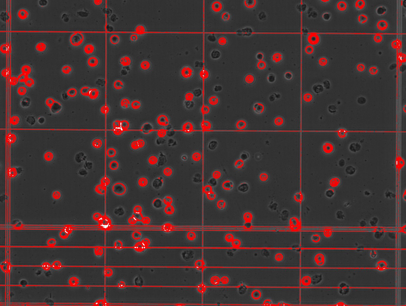
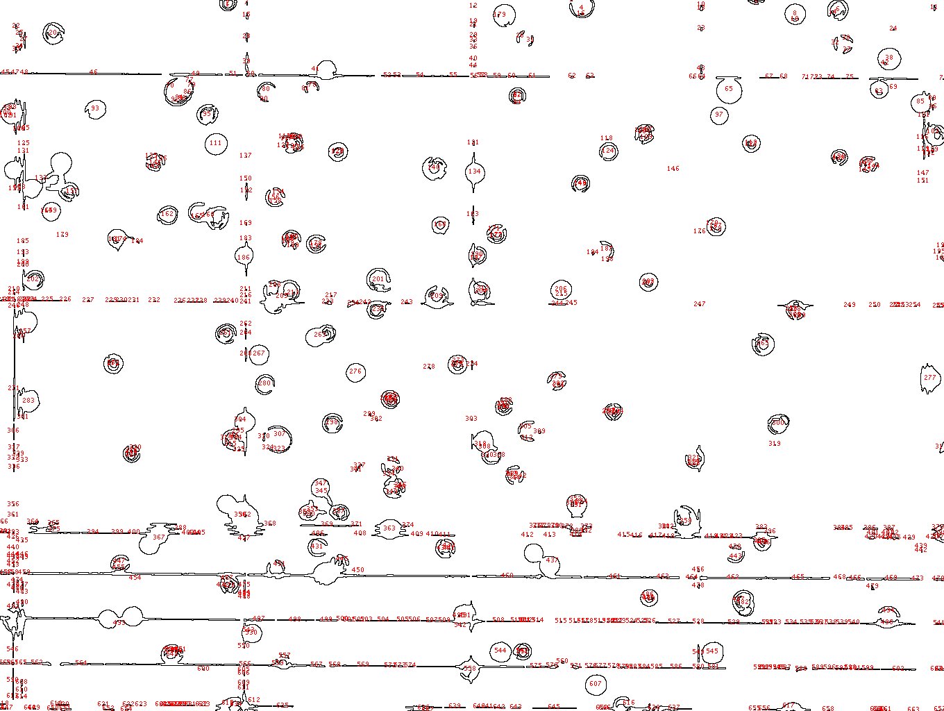
Unfortunately, it is not just about not using the grid. Cell counting chambers are specifically made to count cells in a volume (As in, there is a precise separation between the coverslip and the chamber - Usually 0.1mm) which allows you to do the counting accurately, even if you pipet a semi-variable amount each time. So if you want to use automatic methods, you still need a cell counting chamber-like device. It's not enough to put liquid on a glass slide and put a coverslip on it, the volumes would not match and probably be much less accurate.

Even if you manage to get a counting chamber-like device without the grid, you still need to keep the stereology counting rules like for the manual counting (Keep objects if they intersect the bottom and left corners, discard if they are in contact with the upper and left corners) in your counting pipeline, or you rist over/under estimating the amount by a fair percentage.

All in all, you risk having a less accurate count by doing it automatically  than if you counted a few chambers manually... And loose more time in the acquisition and processing then if you had just counted them by hand.

Best

Oli


Hi Tizy,

as far as I can remember something like this has been on the list a while ago - the final solution was simply to avoid the grid.  You need to calibrate the scale of your image separately (with your grid or any other object of known size) and keep the magnification.

Michael
________________________________________________________________
On May 17, 2012, at 21:20, tizianarossetti wrote:

> I'm having a similar issue, the resolution in my case is fine, I can visible
> discriminate bitten live and death cells as showed in the link
>
> http://imagej.1557.n6.nabble.com/file/n4992111/cells_viability.jpg
>
> the problem is with the analysis I have many quantification of the
> background that shouldn't be there.
>
> http://imagej.1557.n6.nabble.com/file/n4992111/cells_counting_analysis.jpg
>
>
> any suggestion?
>
> Cheers
>
> Tizy