Hey folks,
maybe one of you guys has an idea how to solve my problem - I want to combine serial angiography pictures of an aneurysm into one single image...
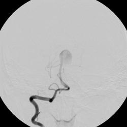
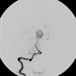
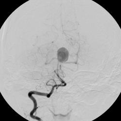
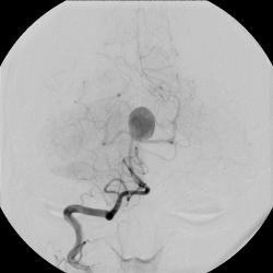
so this is how a basilar tip aneurysm looks like (yes, the round sac). As you can see there is quite a contrast medium flow dynamic inside the aneurysm sac (first it is circular, then more diffuse - I left out a couple of slices, usually you can see the dynamics even better) and the gray scale values of different points vary over time. Now i want to compile a single image that shows me, where the most contrast medium is over a certain time period, i.e. where is the most "flow" inside the aneurysm sac.
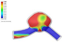
The final goal is to take this single compiled picture and perform a color coding to get something that looks simialr to this (although a complete different technique) --->
Maybe one of you has ma clue?
Thanks a lot and Greetings, Mat

