Dear Seb,
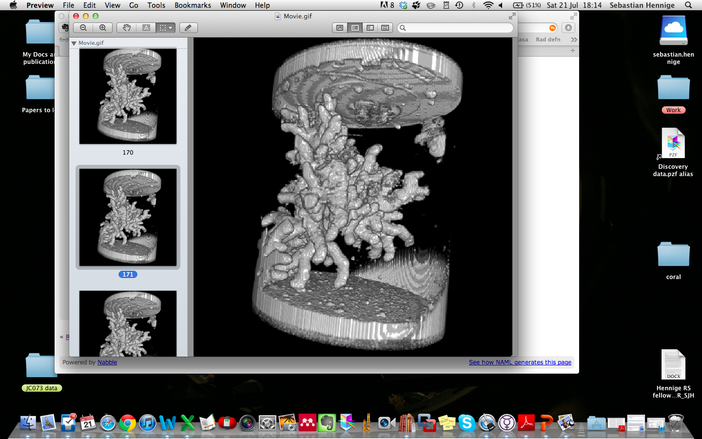
Would it be possible for you to create a circular ROI around the coral, then
"Edit>Clear Outside" (all slices) to remove the tube before reconstructing?
That would reduce the problem to differentiating between the coral and
water. As Michael has said that would be much easier on CT than MRI.
I often meet similar issues when 3D reconstructing CT scans of the pelvis.
There are typically metal structures (stretcher, external fixators,
monitoring equipment etc) that lie around the patient and need to be
isolated from the bone to get a decent reconstruction-the "clear outside"
method works very well for that.
Kind regards,
Julian
Julian Cooper
Consultant Orthopaedic Trauma Surgeon
Birmingham UK
-----Original Message-----
From: ImageJ Interest Group [mailto:
[hidden email]] On Behalf Of sjhenn
Sent: 19 July 2012 21:11
To:
[hidden email]
Subject: Reconstructing a 3D image of an object in water
Dear List,
I have a question which I hope someone can help me with;
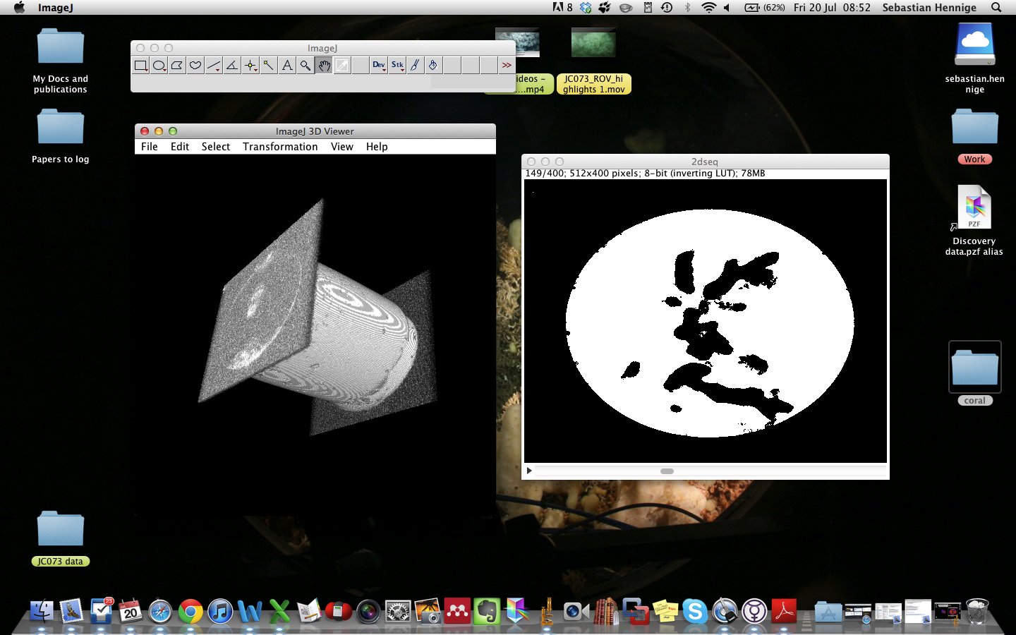
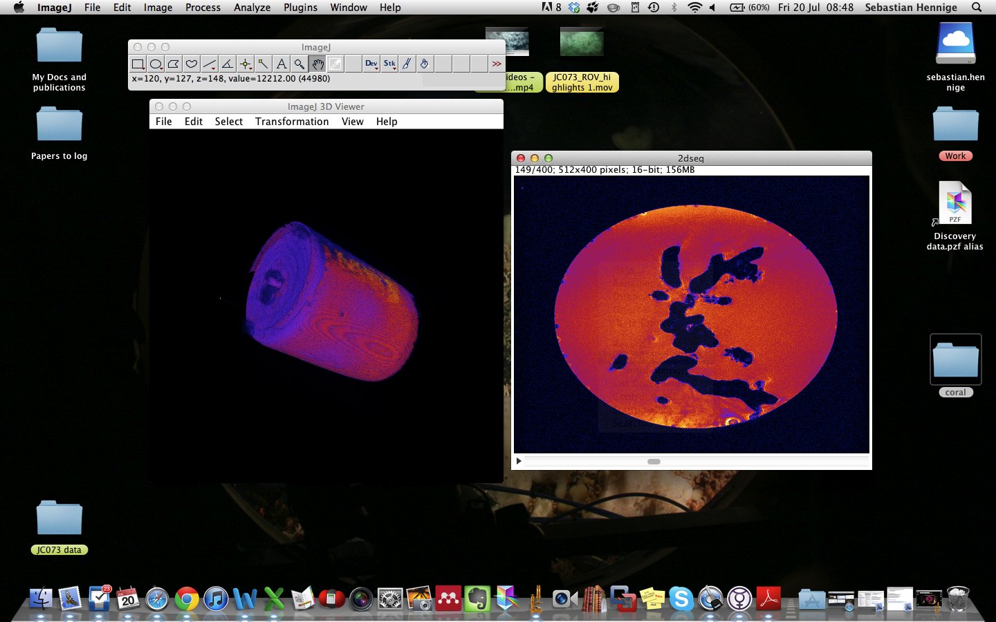
We have an MRI scan of some coral in a tube full of seawater. We are trying
to image this coral in 3D (removing the tube and the water), but even
through the 3D imaging plugins, we can only ever get a 3D image of the tube
that the coral in is. We have tried subtracting the background but this only
got rid of the water within the tube, and not the tube itself. We can
visualise the coral in various ways but seem unable to isolate it. If anyone
has any advice on this it would be greatly appreciated!
Best wishes,
Seb
--
View this message in context:
http://imagej.1557.n6.nabble.com/Reconstructing-a-3D-image-of-an-object-in-water-tp4999503.html
Sent from the ImageJ mailing list archive at Nabble.com.
--
ImageJ mailing list:
http://imagej.nih.gov/ij/list.html--
ImageJ mailing list:
http://imagej.nih.gov/ij/list.html