Hi,
I can recommend "Cell profiler" software (free on-line), also you can try
and image at x60 and do not do z-stack image...for the analysis use only
one plane..
Rona
On Thu, Mar 8, 2012 at 3:37 AM, eroy <
[hidden email]> wrote:
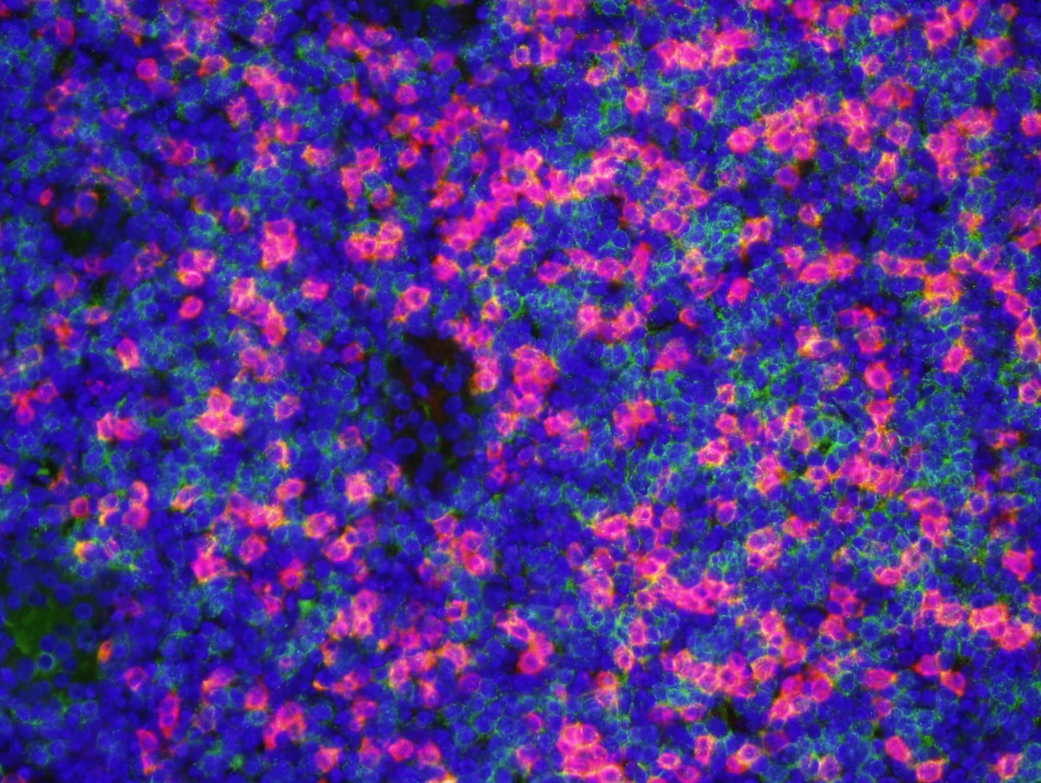
> I have images of T cells in lymph nodes or tumors, stained with an anti-CD8
> antibody and another membrane protein (fluorescent, red or green), so the
> fluorescence generally forms an annulus around the DAPI stained nucleus
> (blue). When the cells are closely packed as in the lymph node, the
> membrane
> fluorescence may be nearly continuous. If I define a cell by its DAPI
> stain,
> I'd like to threshold on an annular region around each nucleus since this
> is
> where the membranes are. The images are typically at 10x-40x. It seems that
> surely this has been done before, but I can't find it. Can anyone direct me
> to a discussion of a similar problem?
> Thanks
> Ed Roy
> University of Illinois at Urbana-Champaign
>
>
http://imagej.1557.n6.nabble.com/file/n4557067/2674-2_Irr_sec2_LN1_40x_red_%5B2%5D.jpg>
> --
> View this message in context:
>
http://imagej.1557.n6.nabble.com/counting-cells-with-a-membrane-marker-tp4557067p4557067.html> Sent from the ImageJ mailing list archive at Nabble.com.
>