Hello,
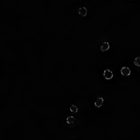
I have numerous fluorescence microscopy images of cell nuclei. A protein in the membrane of the nucleus is labeled with a fluorescent protein and so visualized. I would like to compare/quantify the occurrence of clustering of this protein in a wild type cell versus a mutant cell. I suspect by visual inspection that the distribution of the bright clusters along the roughly circular nuclear membrane differs int he wild type and mutant cells. However, I don't really know of a way to show this in a quantitative manner.
So, my questions are the following:
1) is there an easy way to find clusters in such an image in an unbiased way?
2) is there an easy way to quantify the average brightness of the clusters?
3) is there an easy way to count such clusters on a cell-by-cell basis?
4) is there an easy way to quantify or compare the distribution of such clusters along a more or less circular membrane?
I have attached a sample image of what an image would look like. In this case 7 nuclei are fully visible. Thanks very much in advance for any help that will be offered.
